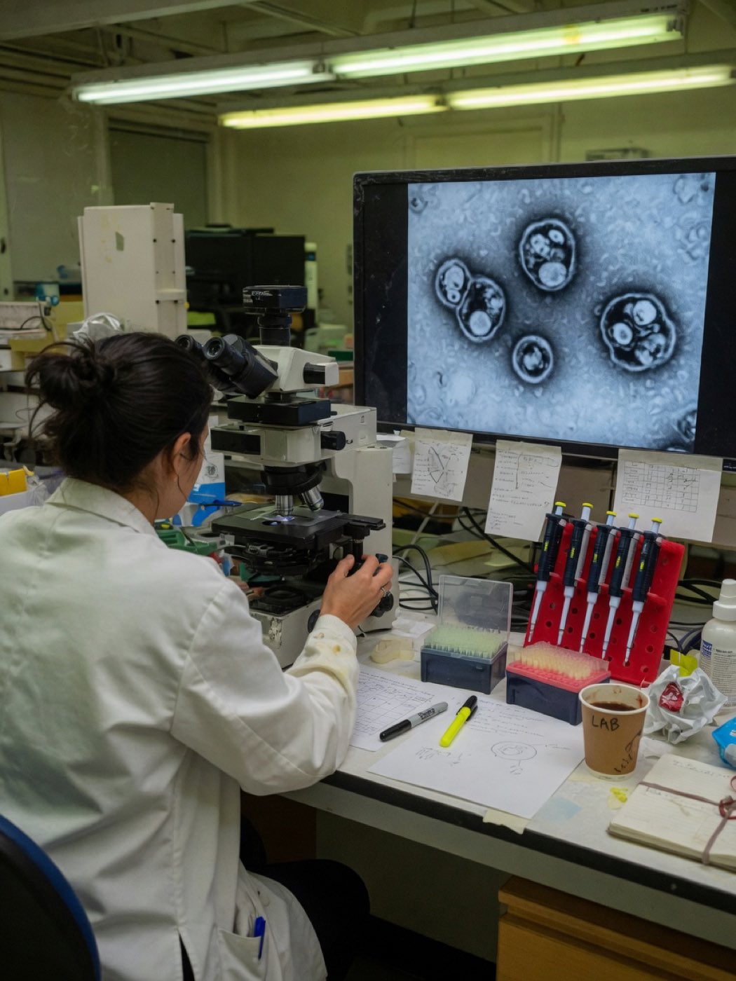
Guided · nine steps

Start here.

No background needed and no vocabulary assumed. Nine short steps, in order, each one a single idea. If you have never heard the word exosome, you are exactly who this was written for. Two of the steps ask you to commit to a number before they show you the answer. Do that part properly — being wrong is the point of it.

Begin

Step 1 of 9

What you will not find here: a price, a booking form, or a sentence about what any of this does for a person. This page describes biology and published research. That is the entire remit.

01 / 09

How small that actually is

Eight hundred of them across one human hair.

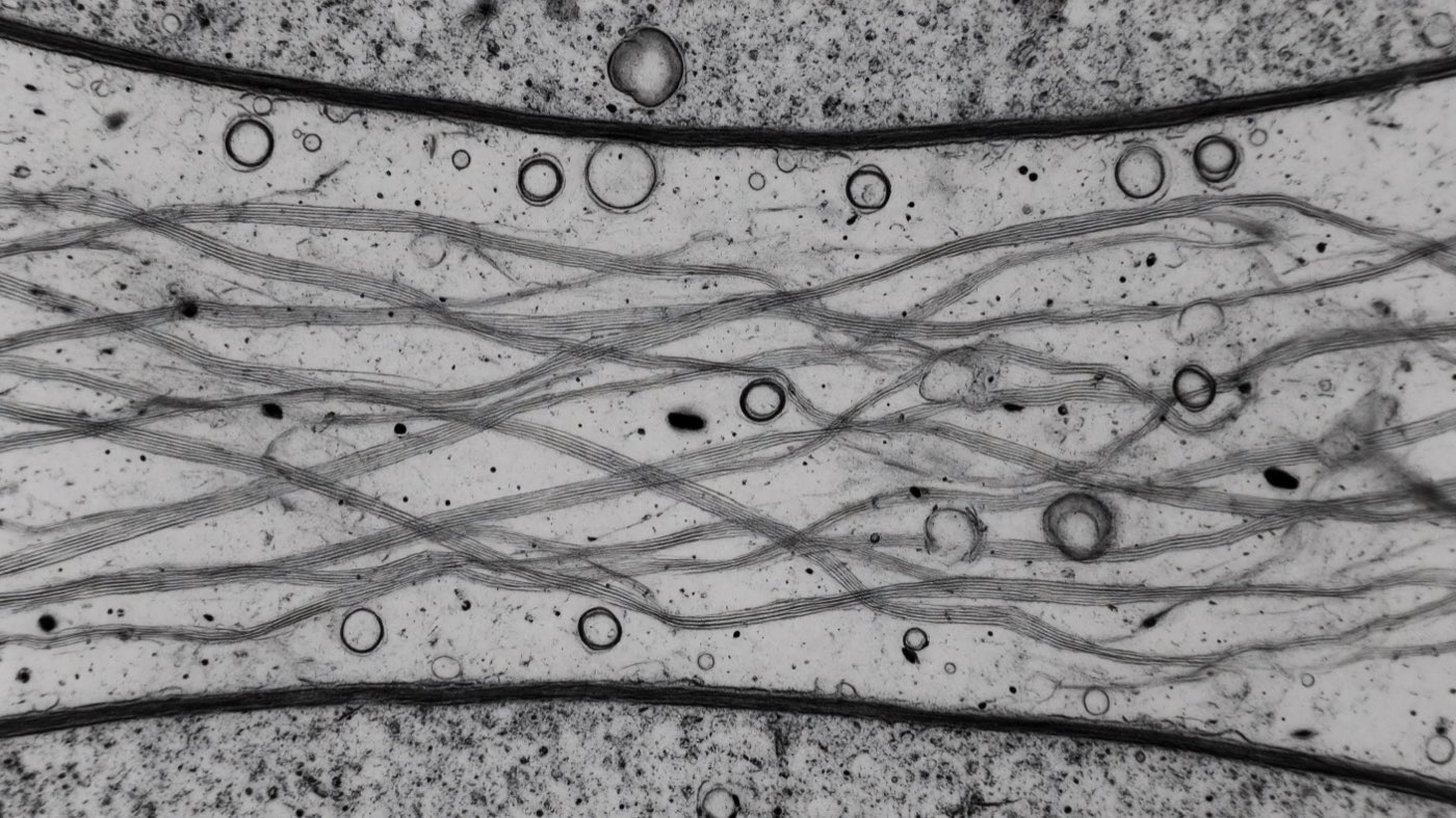
Modelled electron-micrograph view: a narrow gap between two cell surfaces, crossed by bundles of collagen fibres, with vesicles scattered among them at a fraction of their thickness

30–150 nm

Every number on this page came out of an instrument, and the instrument is part of the claim.

Plain language

A human hair is about 80,000 nanometres across. A red blood cell is about 7,000. These vesicles are 30 to 150 — call it 100.

So about eight hundred of them fit across the width of one hair. Ten thousand of them in a line makes one millimetre.

The membrane wrapped around one is roughly five nanometres thick. On a 100-nanometre sphere that is proportionally thinner than the skin of a soap bubble — worth holding onto, because it is the part that has to survive being frozen.

Every comparison in this step is a comparison of size and nothing else. The hair, the blood cell, the soap bubble: analogies for how small, not for what anything does.

Full detail

Why a size is always a range and a method. Nanoparticle tracking analysis infers a diameter from Brownian motion in suspension and reports a hydrodynamic size. Transmission electron microscopy images a fixed, dehydrated, contrast-stained specimen and typically returns a smaller number. NanoFCM measures scattered light against calibrated standards.

In the study specific to this cell line, NanoFCM gave 50–150 nm and TEM gave approximately 80 nm. Same particles, two instruments, two answers. A single headline diameter with no method attached to it is not a measurement.

A geometry check worth doing once: a 100 nm sphere has a volume of about 5.2 × 10⁻¹⁹ litres. Ten billion of them occupy roughly 5.2 nanolitres — about one 191,000th of a one-millilitre fill. Whatever is being counted, it is not filling the container.

Line up 100-nanometre vesicles edge to edge until they span one millimetre. How many does that take?

Show the answer 10000 vesicles — across one millimetre

Ten thousand. One millimetre is one million nanometres, so a 100 nm particle fits ten thousand times across the width of a pencil line. This is the number the glowing-orb render in every other version of this picture quietly hides.

One hundred nanometres. Ten thousand across a millimetre. Hold that number — everything after it is measured against it.

02 / 09

Where they are made

Inside the cell, in a compartment full of smaller bubbles.

Modelled compartment inside a cell, holding dozens of much smaller vesicles

Plain language

Almost every kind of human cell sheds membrane-wrapped packets into the fluid around it. They are not debris. They are one of the ways cells pass material to each other without touching.

Follow one back to where it came from. Deep inside the cell there is a compartment — a bag, if the analogy helps, and it is only an analogy — whose own wall keeps folding inward and pinching off tiny bubbles into the space inside it. Biologists call it a multivesicular body: one bag holding dozens of much smaller ones.

Those small bubbles are built inside the cell and never touch the outside until the whole compartment is emptied out. That matters, because it is the difference between the two things a jar of these particles can contain: bubbles built inside a compartment, and bubbles pinched straight off the cell’s outer skin. Same size range, different origin, and no ordinary method can tell them apart afterwards.

Full detail

Nomenclature. “Exosome” properly denotes biogenesis rather than size: intraluminal vesicles that form inside a multivesicular body and are released when that body fuses with the plasma membrane. Most preparations cannot demonstrate that origin, so the field’s reporting guidelines prefer small extracellular vesicle for anything defined by size and density alone. A site that uses the two words interchangeably is, in strict terms, describing a size fraction.

Extracellular vesicles (EVs) are released by most cell types. Small EVs in the 30–150 nm range originate in the endosomal system: inward budding of the limiting membrane of a late endosome produces intraluminal vesicles, and fusion of that multivesicular body with the plasma membrane releases them.

Conventional surface markers are the tetraspanins CD9, CD63 and CD81. The vesicles characterised in the one published study specific to this cell line were positive for all three.

Two particles, both about 100 nanometres, both wrapped in membrane. One formed inside a compartment and left when that compartment fused with the cell’s outer membrane. The other was pinched straight off the outer membrane. Which one does the word exosome strictly refer to?

  • The one formed inside a compartment Correct

    That is the strict definition: an exosome is an intraluminal vesicle from a multivesicular body. The word names an origin, not a size.

  • The one pinched off the outer membrane

    That one has its own name — a microvesicle, or ectosome. It can be exactly the same size, which is precisely why size alone cannot settle the question.

  • Both — the word just means a small vesicle

    That is how the word is used in practice, and it is why the field’s own reporting guidelines prefer small extracellular vesicle for anything defined by size and density alone.

Nothing later on this page depends on winning that distinction, but it is worth knowing that the two commonest words in this field name different things and get used interchangeably anyway. A preparation defined by size and density is a size fraction. Calling it an exosome is a claim about where it came from, and most preparations cannot demonstrate that.

Built inside a compartment, not shed off the surface. The word exosome names an origin — and most preparations cannot demonstrate it.

03 / 09

What goes inside

Whatever happens to be there when the wall folds in.

Labelled cross-section of an extracellular vesicle A cut-open vesicle, described from the outside inward: a glycocalyx of surface glycans; membrane-associated proteins resting on the outer leaflet; tetraspanins CD9, CD63 and CD81 passing four times through the membrane; the phospholipid bilayer of two opposed leaflets; the enclosed aqueous lumen; and three luminal cargo classes drawn as classes rather than named species — soluble protein, mRNA and miRNA. Glycocalyx Membrane-associated proteins Tetraspanins — CD9, CD63, CD81 Phospholipid bilayer Lumen Soluble protein cargo mRNA cargo miRNA cargo 01 02 03 04 05 06 07 08 SCHEMATIC · NOT TO SCALE · 30–150 NM CLASS
Schematic cross-section, not to scale: membrane thickness is exaggerated against particle diameter so the two leaflets can be told apart. Luminal contents are drawn as classes, not as an identified inventory.

≈5 nm

The thickness of the membrane. One twentieth of the diameter it encloses.

Plain language

Nothing loads these. There is no packer. The compartment wall folds inward, and whatever happens to be against that patch of wall — molecules stuck to it, molecules floating just inside it — is what gets sealed in when the bubble closes.

Some of what is inside is not cargo at all. The sorting machinery that does the folding gets caught in its own parcel, and that turns out to be useful: finding those particular proteins inside a vesicle is one of the few ways to argue it came from a compartment rather than off the cell’s surface.

There is also a hard limit set by geometry. At about 100 nanometres across, a great deal simply does not fit. A single mitochondrion is 500 to 1,000 nanometres — five to ten times the width of the whole vesicle. The nucleus is far larger again. Whatever is in there is small, and there is not much room.

Full detail

ALIX, TSG101, syntenin-1 and flotillin-1 are components of the endosomal sorting machinery that buds a vesicle inward off the wall of a multivesicular body. They end up inside the vesicle because they were part of making it — which is exactly why their presence is used as evidence of endosomal origin.

The protein fraction. Part of it is the machinery that built the vesicle in the first place and therefore travels with it. The rest is the enriched category found by proteomics on this cell source: proteins annotated to cell adhesion and extracellular matrix organisation.

The bilayer is not inert packaging. Its composition is measurably different from that of other vesicle subclasses released by the very same cells, and one of its lipids is read directly by the immune system as a recognition signal.

Take it apart. Three layers, from the outside in.

  • The membrane

    A lipid bilayer about 5 nanometres thick. On a 100-nanometre sphere that is one to twenty across the diameter — proportionally thinner than the wall of a soap bubble. It looks wrong the first time you section it. It is not wrong; that is the proportion.

  • The surface proteins

    Tetraspanins — CD9, CD63 and CD81 — sit in the membrane and protrude from it. They are the panel used to argue that a preparation is what it says it is, and the vesicles in the one published study specific to this cell line were positive for all three.

  • The inside

    The lumen: a few attolitres of the cell’s own fluid, plus whatever was sealed in with it. Small RNA, soluble protein, some of the machinery that built the parcel. Step 7 counts what is actually in there, and the count is the surprise.

That is the whole object: a bilayer, a set of surface proteins, and a very small interior. No motor, no compass, no timer. Everything that happens to it downstream is a consequence of those three things and of the fluid it lands in.

Cargo is enclosed, not loaded. And the machinery that built the parcel travels inside it, which is how its origin gets argued.

04 / 09

The way out

The membrane opens outward. Almost everyone pictures it backwards.

Modelled membrane fusion: an open crater whose interior is continuous with the space outside the cell

Plain language

The compartment travels to the edge of the cell and its wall meets the cell’s own outer wall. The two join. For a moment there is no boundary at all between the inside of the compartment and the world outside the cell — one continuous space, an open crater rather than a closed pocket.

The small bubbles are now outside. Notice that they never crossed a membrane to get there. Nothing was pushed through anything; a wall was removed.

Run that same picture backwards and you get a completely different process: a cell closing a pocket around something and drawing it in. That is what happens at the far end of this journey, in step 6. Same shapes, opposite direction — and getting the direction the wrong way round is the commonest mistake made about these particles, including by the software that draws them.

Full detail

The event is membrane fusion. The limiting membrane of the multivesicular body fuses with the plasma membrane, the lumen of the compartment becomes continuous with the extracellular space, and the intraluminal vesicles are released without ever traversing a bilayer. Once released, they are what the word exosome denotes.

The mirror-image process — inward invagination of the plasma membrane, enclosing extracellular material inside a new intracellular compartment — is endocytosis. Exocytosis and endocytosis are not variants of one another: they differ in the direction the membrane moves and in what ends up enclosed.

The sequence on this step is a model, drawn to show the geometry. It is not a micrograph and no frame of it is data. Where this page shows a measurement it names the instrument that produced it.

You are watching the release. Which way does the membrane move?

  • Outward — the inside of the compartment opens into the space outside the cell Correct

    Yes. The two membranes fuse and the lumen becomes continuous with the outside. There is no closed circle anywhere in the correct picture.

  • Inward — the cell closes a pocket around the vesicles

    That is the opposite process, and it is the mistake worth making once. A closed pocket drawn inward is endocytosis — a cell taking material in. Step 6 is that.

  • Through — the vesicles pass out through the membrane

    Nothing of this size crosses an intact bilayer. If a drawing shows a sealed sphere on the far side of an unbroken membrane, the drawing has skipped the only event that matters.

Outward. It is worth being pedantic about, because the direction is the mechanism: the compartment is emptied by having its wall removed, not by pushing anything through one. The clip on this step was checked frame by frame for exactly that — the interior of the crater has to be one continuous space with the outside, with no closed circle anywhere.

Release is a wall being removed, not a parcel being pushed through one. Outward at this end of the journey; inward at the other.

05 / 09

Out in the crowd

No engine, no steering, no destination.

Modelled extracellular channel crossed by bundles of collagen fibres, with vesicles drifting among them

≈30 min

Reported circulating half-life in a mouse model. A rodent figure, and it is labelled as one.

Plain language

Outside the cell it is crowded and wet: water, salts, sugars, loose protein, and the fibrous mesh that cells sit in. Into that goes a 100-nanometre bubble with no engine, no propeller and no sense of direction.

What moves it is collision. Water molecules strike it from every side, unevenly, millions of times a second, and it jitters. That jitter is called Brownian motion, and it is the only transport it has. Push a whole population in one direction and the push is erased in about two seconds — thermal collision beats any steering force by orders of magnitude and does not care how hard you push. Distance is not the problem out here. Direction is, and there isn’t any.

Where vesicles have been tracked in the bloodstream of laboratory animals, they disappear quickly. Most of the signal is gone within about half an hour, taken up by scavenger immune cells concentrated in the spleen and liver.

Full detail

At roughly 100 nanometres in aqueous medium, transport is diffusive. The Stokes–Einstein relation puts the diffusion coefficient of such a particle in water at the order of a few square micrometres per second — fast across a single cell, negligible across a tissue. Directed motion requires a force that exceeds thermal noise, and nothing about a passive vesicle supplies one.

In a mouse model, reported circulating half-life is on the order of 30 minutes, with roughly a 200-fold reduction in signal by 30 minutes and dominant accumulation in spleen and liver. Clearance is primarily phagocytic, mediated by macrophages of the reticuloendothelial system.

Rodent biodistribution is rodent biodistribution. There are no published human pharmacokinetic data for vesicles from this cell source.

Tap the thing that decides where one of these ends up.

  • A signal inside it, telling it where to go

    There is nothing in there that could. No motor, no energy supply, no receiver. Whatever is in the lumen is cargo, not a navigation system.

  • An address on its surface that picks a destination

    Surface molecules do matter — they decide which cells it can dock with once it is already touching one. That is a handshake, not a homing system, and no targeting or homing behaviour is established for material from this cell source.

  • The random motion of the fluid, plus wherever that fluid is going Correct

    That is it. Thermal collision over short distances, bulk flow for anything further, and in an animal, the phagocytes of the spleen and liver for most of the population within about half an hour.

This is the least glamorous step on the page and the most load-bearing. A particle with no steering goes where the fluid takes it, which means where it is put decides almost everything about where it can arrive — and that is why a result measured in a dish is a result about a dish.

It does not travel. It is carried. Where a particle is put decides what it can reach, which is why the system a result came from is part of the result.

06 / 09

Getting inside is the easy part

And then it is inside a bag, inside the cell.

Modelled uptake: a narrow-necked cup of membrane closing around a vesicle

Plain language

A vesicle drifts into a cell and the molecules on its outer surface act like a handshake, testing whether this is a cell worth interacting with.

The cell swallows the vesicle into an internal bubble called an endosome. That sounds like arrival. It is not. By default that bubble is on its way to the lysosome — the cell’s recycling compartment — where everything inside it is broken down.

For anything inside the vesicle to matter, it has to escape that internal bubble before it is digested. Most of it does not. Published estimates of successful escape are typically low single-digit percentages.

This is the biggest unsolved problem in the entire field, and it is why a number printed on a label is so difficult to interpret — the great majority of what is counted never functionally arrives anywhere.

Full detail

Internalisation proceeds through clathrin-mediated endocytosis, caveolin-dependent endocytosis, macropinocytosis, phagocytosis, lipid-raft-dependent routes and, less commonly, direct fusion with the plasma membrane. Which route dominates is cell-type and context dependent.

Internalised vesicles traffic to early and then late endosomes. The default destination is lysosomal degradation.

Functional delivery requires endosomal escape: back-fusion of the vesicle membrane with the limiting endosomal membrane, releasing luminal contents into the cytosol. The process is inefficient and is the recognised rate-limiting step for all vesicle-based delivery.

Escape efficiency is not measured on release panels. Nobody reports it per lot, because there is no standard assay for it.

The membrane has closed around it and it is inside the cell now, in a small sealed compartment. Has the cargo arrived?

  • Yes — inside the cell is inside the cell

    Not yet. There is still a membrane between the cargo and the cell’s interior, and by default that compartment is on its way to be broken down.

  • No — it is inside a bag inside the cell, and the bag is heading for the recycling compartment Correct

    Correct, and this is the unsolved problem of the whole field. The cargo has to get out of the endosome before the endosome acidifies. Published estimates of successful escape are typically low single-digit percentages.

  • Only if the cell was expecting it

    Uptake is not gated on being expected. Cells internalise material continuously, and being taken in is not a sign of being wanted.

Getting inside is routine. Getting out of the compartment that carried you in, before it turns acidic, is the recognised rate-limiting step for every vesicle-based approach anyone has built — and it is not measured on any release panel, because there is no standard assay for it.

Uptake is release run backwards: the membrane moves inward here. And uptake is not arrival — most of what gets in is digested.

07 / 09

What is actually in the envelope

A gene cluster only primates have.

Giemsa-banded metaphase chromosome spread

1 : 121

Measured copies of a given microRNA per vesicle, averaged across six sources: roughly one copy for every 121 vesicles.

Plain language

Cargo lists in this category all read the same: microRNA, proteins, lipids. True, and close to useless, because the interesting questions are which ones and how many.

One answer is specific to the placenta. There is a cluster of microRNA genes on human chromosome 19 — around 59 of them — that is switched off in essentially every human tissue except the placenta and very early embryonic cells. It exists only in primates. Mice do not have it at all.

Which means a mouse study cannot model this part of the biology. Not models it imperfectly. Cannot.

Here is the mental model nearly everyone arrives with: each vesicle is a tiny loaded syringe, packed with instructions, docking with a cell and delivering them.

It is wrong, and it is wrong by about two orders of magnitude. Before you read on, put a number down.

Full detail

C19MC — the chromosome 19 microRNA cluster: 46 genes, roughly 59 mature microRNAs, spanning about 100 kb at 19q13.41, imprinted and expressed from the paternal allele, and epigenetically silenced outside placenta and embryonic stem cells.

Note what that does to the preclinical literature in this area. Rodent work cannot be modelling a mechanism the rodent genome does not encode, so any inference drawn from it about this particular cargo is an inference about something else.

And a cargo list is not a specification. No standardised assay relates the composition of a preparation to any measurable biological activity, so “contains microRNA” on a website carries no quantitative content whatsoever.

Chevillet and colleagues quantified microRNA stoichiometry across six sources using absolute quantification against synthetic standards. The mean was 0.00825 molecules of a given microRNA per exosome. Even for the most abundant microRNA in the most concentrated preparation, occupancy stayed far below one copy per vesicle.

Stack that on the endosomal escape problem — cargo that is taken up still has to leave the endosome before it is degraded, and typically only low single-digit percentages do — and you can derive the field’s central limitation for yourself. The arithmetic of delivery does not currently work, which is a large part of why this area is still preclinical.

How many vesicles would you have to open to find one copy? Pick a number before you read on. You are opening extracellular vesicles one at a time, looking for a single copy of one abundant microRNA — the kind of molecule every site in this field says these particles carry.

Show the answer 121 vesicles — about 121 vesicles per copy

Across six different sources, the measured mean was 0.00825 copies of a given microRNA per vesicle. That is roughly one copy for every 121 vesicles. So most of them are carrying none of it at all. A vesicle is not a tiny syringe. Whatever these particles do, they do it as a population, not as individual couriers — which is also why nobody in this field has agreed on what a dose even means. Chevillet et al., PNAS 2014;111(41):14888–93, PMID 25267620 · six sources.

There is no such thing as one vesicle that does something. If there is an effect anywhere, it belongs to a population and not to a particle.

08 / 09

What one study actually found

And the question you should ask about every result like it.

Researcher analysing cellular imagery at a confocal microscope

Plain language

Cells that stop dividing do not necessarily go quiet. Some enter a state called senescence, in which they keep secreting a set of signalling proteins into their surroundings.

In 2026 a group at the NIH National Institute on Aging, working alongside scientists from the company that developed the cell line, published what happened when trophoblast-stem-cell vesicles were added to senescent human skin cells in a dish. Measured levels of three of those proteins — CXCL1, IL8 and GDF15 — went down. Markers of DNA damage went down. NF-κB signalling was suppressed.

Now the part that matters more than the result. In vitro. Human dermal fibroblasts. One study. No animals. No people.

If you take one habit away from this page, take that one: before you believe any finding in this field, ask in what system?

Full detail

Aging Cell 2026;25(2):e70368. NIA intramural programme, grant AG000511, with five employees of the cell-line developer among the co-authors — which makes it NIH-collaborative, not NIH-independent. The distinction is worth keeping, because the second phrasing gets used loosely across this category.

Vesicle characterisation in the same paper: 50–150 nm by NanoFCM, approximately 80 nm by TEM, positive for CD9, CD63 and CD81.

What the study does not establish: anything about an animal, anything about a person, any route of administration, any dose, and any durability beyond the culture period.

The published study this step is built on is the strongest piece of evidence in this whole project. How many people took part in it?

Show the answer 0 people — no animals, no people

Zero. It was cultured human cells in a dish — no animals and no people. That is not a criticism of the study; it is what the study was. Asking in what system before asking how much is the single most useful habit anyone can carry away from this page.

Measured levels changed in cultured cells, in one study, with company co-authors. That is the finding, and the system it was measured in is part of it.

09 / 09

What is not known

The honest summary, in one place, with nothing softened.

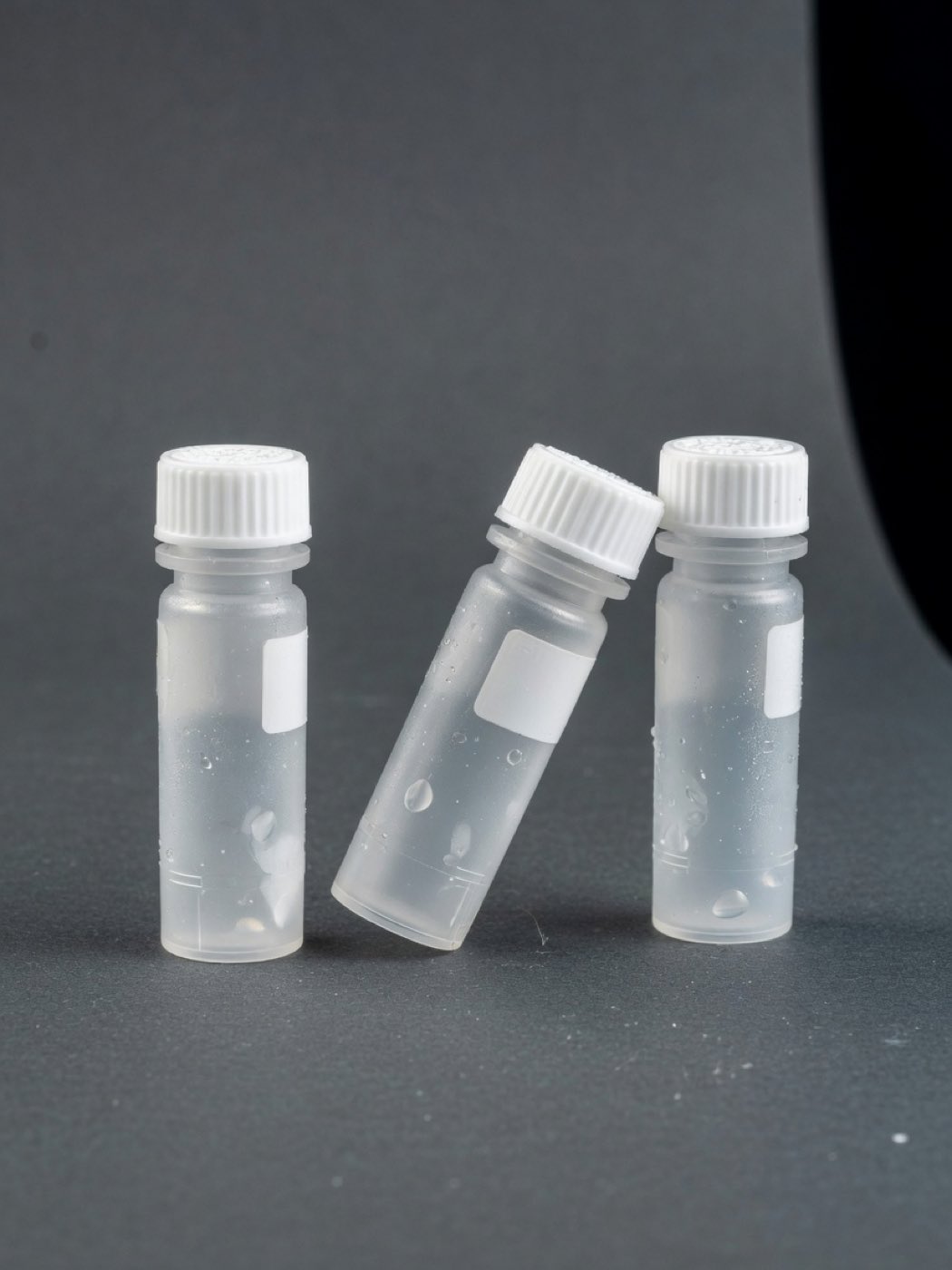
Cryogenic vials of biologic material

Plain language

The state of it, in full:

There are no FDA-approved exosome products. Not for anything.

No clinical trial of trophoblast-derived vesicles is registered anywhere in the world. Zero.

One published study is specific to this cell line, and it was cells in a dish.

Human data elsewhere in the extracellular-vesicle field comes from entirely different cell sources and does not transfer.

No public teratoma or tumorigenicity dataset exists for these lines. What exists is karyotype stability, which is a different question answering a different thing.

The safety panel that does exist is a topical ISO 10993 series — cytotoxicity, irritation, sensitisation, phototoxicity. It says nothing about any other route.

A field can be genuinely interesting and almost entirely unproven at the same time. This one is both, and pretending otherwise is how companies in this category end up in warning letters.

Full detail

Grade: preclinical. In vitro and animal work, one in-vitro publication specific to these vesicles, early-phase human data only in the adjacent mesenchymal-stromal-cell field, no approved product anywhere, and active FDA enforcement in this space.

Borrowing evidence across cell sources is the commonest form of overclaiming here. A phase 3 programme in bone-marrow MSC vesicles is evidence about bone-marrow MSC vesicles.

Quality-control caveats that qualify every number on this page: no standardised assay for biological activity, no agreed dosing unit, substantial variability by culture format and isolation method, and frequent conflation of conditioned medium with purified vesicles.

One thing the reported release panel does not include. HLA-G is real trophoblast biology and it is described in the placental vesicle literature. It is not on the release panel for the material described here, and nothing on this page states that these vesicles carry it. The reported panel is CD9, CD63 and CD81.

The 2012 paper that much of this cell line’s early literature was built on was retracted on 11 February 2021. The grounds were image duplication, missing raw data, underpowered statistics and an undisclosed editorial conflict. It carries a retraction notice on PubMed. This page does not cite it, and the 2015 follow-up explicitly builds on it, so that is not a clean substitute either. You will not find either one anywhere in these nine steps, and that absence is deliberate.

Two trays. On the left, statements that are established — on the record, checkable. On the right, statements that are not established. Sort all six.

  • No exosome product has been approved by the FDA, for anything Established

    Established. It is the FDA’s own sentence: “There are currently no FDA-approved exosome products.” The material described here is an unapproved biologic.

  • One published study characterises vesicles from this cell line and reports what happened to cultured human skin cells Established

    Established. Aging Cell 2026;25(2):e70368. In vitro, one study, with company co-authors — collaborative rather than independent.

  • The karyotype of these cell lines has been characterised as stable Established

    Established, and worth stating precisely. Karyotype stability is a real, checkable characterisation. Note what it is not: it is not tumorigenicity data and it does not answer that question.

  • There is public data ruling out tumour formation for these cell lines Not established

    Not established. No teratoma or tumorigenicity dataset is public for these lines, so nothing rules that out and this page does not claim anything does. What exists is karyotype stability — a real, checkable characterisation that answers a different question.

  • A clinical trial of trophoblast-derived vesicles is under way somewhere in the world Not established

    Not established. No clinical trial of trophoblast-derived vesicles is registered anywhere in the world. Zero. Human data elsewhere in this field comes from entirely different cell sources and does not transfer.

  • There is an agreed dose for this class of material Not established

    Not established. No standardised assay relates any measurable property of a preparation to biological activity, and there is no agreed dosing unit. A particle count is a count.

Three of those are on the record and three are not, and the three that are not are the three this category asserts anyway. A field can be genuinely interesting and almost entirely unproven at the same time. This one is both. There is no approved exosome product, the material described here is an unapproved biologic, and enforcement in this area is active — which is exactly why the honest version of this page is the only version worth publishing.

Preclinical. One in-vitro study specific to these vesicles, no registered trial anywhere, no approved product anywhere. Interesting is not the same as proven.
Missions

Missions

  1. 01

    Calibration

    Establish the scale before you trust anything measured on it.

    You crossed seven orders of magnitude and cut the vesicle open. The membrane you saw is the real proportion, not a drawn one.

    01 · 03

  2. 02

    The Checkpoint

    Call which way the membrane moves before the model shows you.

    You committed to a direction and then watched the membrane take it. Outward is release. Inward is uptake, and it is the mistake almost everyone makes.

    02 · 04

  3. 03

    Provenance

    Trace the cargo back to a genome, and commit a figure before the count is shown.

    You put a number on how much cargo a vesicle carries, and traced one cargo class to a gene cluster only primates have. Both of those are facts about the parcel, not about a person.

    07

  4. 04

    The Quiet

    Read the one published result in the system it was run in.

    You read one published in-vitro result with its system attached. That is the whole finding — cells in a dish, one study, several authors employed by the supplier.

    08

  5. 05

    Transit

    Follow the population out of one cell and into another. Every step costs.

    You followed a population from open fluid to the inside of an endosome. Nothing steered, and most of what got in was digested.

    05 · 06

  6. 06

    Audit

    Check the working. Sort what is established from what is not.

    You audited the evidence. Three statements on the record, three not — and the three that are not are the three this category asserts anyway.

    09

Rank
  1. Unranked

    Nothing on the record yet.

  2. Observer

    You put a number on the record before you were shown one.

    1 committed

  3. Recorder

    A mission closed, with every objective in it logged.

    1 missions · 1 committed

  4. Calibrator

    Half the campaign closed, with at least one figure landing close enough to be calibration rather than luck.

    3 missions · 2 committed · 1 close

  5. Metrologist

    Five missions, four figures, and the first of the underlying data opened — you have started checking what the figures rest on.

    5 missions · 4 committed · 2 close · 1 at full detail

  6. Referee

    The whole campaign, every figure committed, and the evidence read. This is the rank this page is actually about: the last thing you did was check somebody else’s working.

    6 missions · 5 committed · 3 close · 3 at full detail

That is the whole mechanism.

Nine steps, and you now know more about how these particles work than most of this category publishes. What you have not been told is what any of it does for a person, because that has not been established and this page will not pretend otherwise. Two doors from here. The science page follows each piece of cargo down to the finding and the reference it came from. The clinic page describes the material, its paperwork and its limits to the people who would handle it.

  • Held on this device. No account, nothing sent anywhere.
  • No percentile and no ranking against other readers. There is no record of other readers here to compute one from.
  • Every scene is a model of biology, not a statement about a product.
This page describes biology and published research. It is not medical advice and not an offer of treatment. The material described is an unapproved biologic: no exosome product has been approved by the U.S. Food and Drug Administration for any use, and nothing here has been evaluated by the FDA. Research described is preclinical — in vitro or in animals — and preclinical findings do not establish benefit in people. Permitted use and classification differ by jurisdiction.